About Ligneous Conjunctivitis (LC)
Ligneous conjunctivitis (LC) is a rare, chronic, and recurrent form of conjunctivitis characterized by the formation of fibrinous pseudomembranes on the palpebral conjunctivae. It is the most common clinical manifestation of Plasminogen Deficiency Type 1 (PLGD-1), an autosomal recessive disorder that leads to reduced plasminogen activity and antigen levels. As a result, there is not enough active Plasmin generated to degrade Fibrin deposits. Ultimately, this deficiency may lead to the formation of fibrinous lesions on mucous membranes throughout the body.
PLGD-1 is a multisystem disorder, and LC may occur alongside systemic fibrinous lesions affecting the respiratory tract, oropharynx, female reproductive tract, gingiva, middle ear, renal collecting system, skin, and central nervous system.
Signs and Symptoms
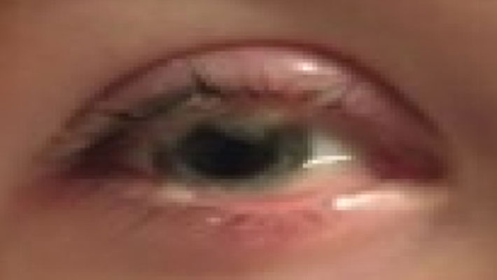
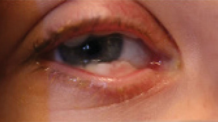
The earliest signs of LC include mucoid discharge, tearing, and redness of the conjunctivae. These symptoms progress to the formation of pseudomembranes on the palpebral conjunctiva, eventually leading to mucosal thickening with a wood-like consistency that replaces the normal eyelid mucosa. Approximately 50% of cases are bilateral, with the upper eyelid most commonly affected, followed by the lower eyelid and bulbar conjunctiva. Patients may experience multiple recurrences or chronic pseudomembranous conjunctivitis, which can precede or coincide with systemic signs such as fever, upper respiratory tract infections, ear infections, or, in females, urogenital tract infections.
- Watery, stringy, pseudomembranous mass
- Accompanied by chronic tearing, redness, and mucoid discharge
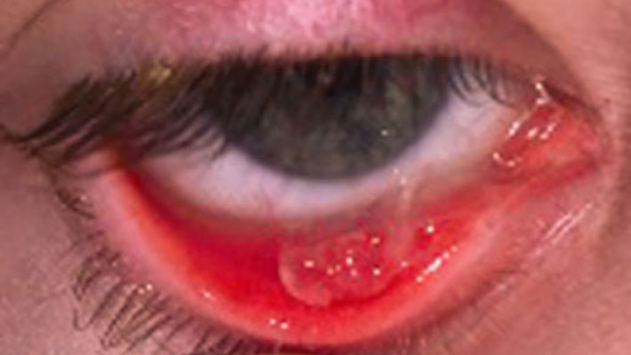
- Followed by palpebral conjunctival pseudomembrane formation
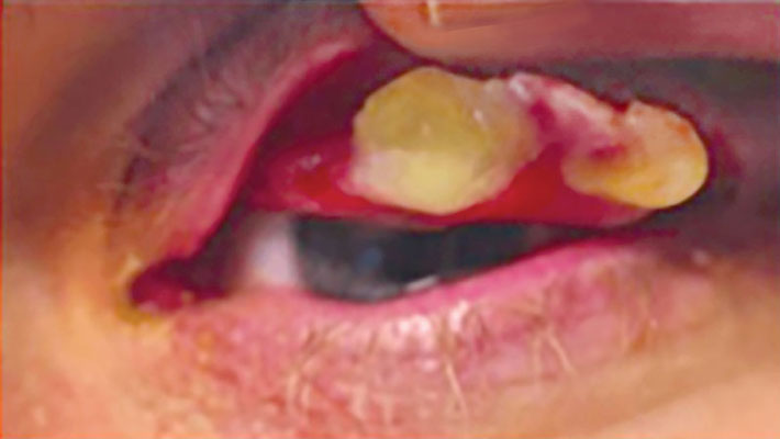
- Progresses to wood-like lesions on the conjunctivae
- Thickened, white, avascular mass
- Watery, stringy, pseudomembranous mass
- Accompanied by chronic tearing, redness, and mucoid discharge
- Followed by palpebral conjunctival pseudomembrane formation
- Progresses to wood-like lesions on the conjunctivae
- Thickened, white, avascular mass
Diagnosis
The initial diagnosis of LC is often clinical, based on patient history and an eye examination. Confirmation typically involves laboratory data showing decreased plasminogen activity and pathological findings of fibrin-rich pseudomembranes.
The diagnosis of ligneous conjunctivitis can be challenging given the pseudomembranous presentation that can be associated with other diagnoses, including viral conjunctivitis, bacterial conjunctivitis, toxic conjunctivitis, allergic or vernal conjunctivitis, as well as amyloidosis. However, recurrence or systemic mucosal involvement—especially in a child—should heighten suspicion. Diagnosis is often definitive when another mucosal site is involved or there is a positive family history.
Treatment
Systemic plasminogen replacement therapy can effectively control conjunctival membranes and other systemic ligneous manifestations. It is approved as safe and effective for the treatment and prevention of the fibrinous lesions associated with PLGD-1 with long-term administration.
Various nonspecific therapies and interventions, including surgery, have been explored in small studies using individual or combination treatment approaches. However, these methods have generally demonstrated suboptimal outcomes. Case reports describing the use of topical or systemic fresh frozen plasma, heparin, corticosteroids, cyclosporine, azathioprine, hyaluronidase, α-chymotrypsin, oral contraceptives, warfarin, and amniotic membrane placement have been published showing inconsistent success.
Surgical excision as a primary intervention can exacerbate inflammation and perpetuate the formation of pseudomembranes due to the trauma induced in these patients, who have impaired wound healing. There is the risk that from excision to excision to time for recurrence of lesions may shorten. Recurrence of pseudomembranes may occur as early as 48 hours after surgery.
Prognosis
The prognosis for LC depends on the ability to control inflammation and prevent recurrence of pseudomembranes. Approximately one-third of individuals with PLGD-1 experience corneal involvement, which can lead to blindness. If left untreated, plasminogen deficiency may result in significant reduction in quality of life and morbidity with potential life-threatening complications.